enesMon - Sat 08:00 - 16:00 +1-713-797-1919
Upload Physician Order
Video Tutorial
Vue Motion
Carestream Portal Access
Portal Access
Physician & Patient Portal Access

ARTHROGRAM

INTRODUCTION

ARTHROGRAM

An arthrogram is an x-ray procedure in which x-ray dye (iodinated contrast) is injected into a joint (shoulder, elbow, wrist, knee, ankle).


This allows the radiologist to identify normal structures, loose bodies, abnormal growths, as well as damage to ligaments and the joint capsule.


The arthrogram is frequently performed in conjunction with a post-arthrogram MRI (magnetic resonance image).

PREPARATION

Do not eat or drink anything at least two hours prior to the exam.

On the day of your exam, please go directly to the 3310 Richmond location. Please be sure to bring the signed order from your physician requesting the examination, and your insurance cards as well.

PRECAUTIONS

You will receive iodinated x-ray dye. If there is a history of an iodinated contrast allergy, the risk-benefit ratio should be discussed with your physician.

Premedication with steroids and/or Benadryl® is generally protective in patients with a history of mild-moderate prior reactions. It is possible to perform an MRI-arthrogram without the use of x-ray dye in some instances.

EXAM

– In the Radiology Department on the day of your x-ray, you will be asked to sign an informed consent form.

– You will lie on an x-ray table. A scout x-ray will be take.

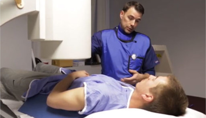
– The technologist will carefully prep and drape the area to be injected. Scout x-ray films will be obtained.

– The radiologist will, with fluoroscopic assistance, inject a local anesthetic. A small needle will be advanced into the joint and contrast injected. A paramagnetic agent is added to the solution if an MRI is to be performed.

– Multiple x-rays will be taken by the radiologist or assistant. This examination takes an average of 15 minutes to complete.

– The radiologist will send a report to your doctor.

;