enesMon - Sat 08:00 - 16:00 +1-713-797-1919
Upload Physician Order
Video Tutorial
Vue Motion
Carestream Portal Access
Portal Access
Physician & Patient Portal Access

DIGITAL MAMMOGRAPHY WITH TOMOSYNTHESIS

INTRODUCTION

DIGITAL MAMMOGRAPHY WITH TOMOSYNTHESIS

Mammography, along with monthly self-examinations and regular physical examinations of the breasts, provides the best chance for early detection of breast cancer. By detecting small tumors before they can be felt and before they have spread, the possibility of a cure is very high.


Early detection makes it possible to give treatment and save the breast. Screening mammography is the key to early detection. This is the only way to find breast cancer before it can be felt. Having routine mammography in conjunction with physical examination is a vital part of your ongoing good breast health.


In breast cancer care, every woman places her trust in accurate and early detection. To live up to this trust, you need to be able to make confident diagnostic decisions. But can you really trust in what you see?  

Increase your diagnostic confidence efficiently and easily with High Definition Breast Tomosynthesis. Already superior with the widest angle, the highest number of projections, and full detector readout, it is now the world’s first tomosynthesis to incorporate EMPIRE Technology:


– See tissue and lesions with unprecedented clarity
– View microcalcifications clearly, and their morphology precisely
– Get Insight, the first synthetic visualization of tomosynthesis in 2D and 3D
– Reduce patient dose by replacing additional mammograms with Insight 2D
– Gain new depth with Insight 3D, for you, your peers and your patients


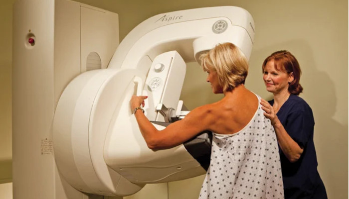
HMI utilizes state-of-the-art digital mammography, shown to be more accurate than film screen mammography among women under the age of 50 years, women with heterogeneously dense or extremely dense breasts on mammography, and premenopausal or perimenopausal women.

Digital mammography offers other advantages over film mammography, including easier access to images and computer-assisted diagnosis (R2® Checker); improved means of transmission, retrieval, and storage of images; and a lower average radiation dose without a compromise in diagnostic accuracy.


PREPARATION

Bring (or arrange the mailing of) all previous mammograms done at another facility. Comparison with these old films could be critically important to interpretation of your new mammograms.

Don’t wear deodorant or powder.

It may also be helpful to wear a two-piece outfit.

PRECAUTIONS

The technologist will take a careful history. If there is a history of a bleeding diathesis, please notify the technologist. Any allergy to lidocaine should also be presented to the technologist in order that an alternate medication be used.

Typically no sedation is necessary for this exam. If you feel a need for sedation, please discuss this with the technologist prior to the day of your biopsy.

You will be positioned on the ultrasound table and the radiologist will perform an ultrasound of the breast. The radiologist will cleanse the skin with betadine and inject lidocaine into the skin and deeper tissues. A small skin nick will be made and the Mammotome™ or core needle advanced into the breast. Needle position will be confirmed at ultrasound and several biopsies performed. Please speak up if there is any discomfort.

Immediately after the biopsy, a radiopaque marker will be deployed at the biopsy site; this will confirm the location of the biopsy and assist the surgeon should a lumpectomy later be required.

Post-biopsy specimen radiograph and mammography may be performed to document the procedure.

The needle will be removed and compression applied to the breast. You will be given an ice pack to be used for four hours after the biopsy.

The final pathologic results are typically available within 2-3 days. A report will be delivered to your physician, typically within 24 hours of receiving the biopsy results.

A technologist will contact you after your procedure; please let her know if you have had any problems or complications.

EXAM

Screening Mammogram

– Please fill history forms as completely as possible.

– A technologist will take two or more x-ray films of each breast. Each breast will be positioned and firmly compressed between two clear plastic plates in order to spread out the tissue. This gives a clear picture of the breast with the least amount of radiation. This may be uncomfortable, but rarely painful. Compression lasts a few moments and then is released immediately.

– If you have sensitive breasts, try having your mammogram at a time of the month when your breasts will be least tender. Try to avoid the week right before your period. This will help to lessen discomfort.

– The films will be studied and interpreted by a radiologist. The results will then be reported to your doctor.

– You may be asked to return for additional views after the radiologist sees your mammogram. These extra views can help the radiologist see an area of your breast in better detail. You may also be asked to return in six months for a short term follow up.


Diagnostic Mammogram:

– Regardless of the results of your screening mammogram, you need to talk to your doctor if you notice a change in your breasts. If you have a lump, thickening, swelling, focal pain, nipple discharge or retraction, or if you are recommended to have additional views, you will have a problem solving study called a “diagnostic” mammogram.

– This study includes mammographic views not done in a screening mammogram. An ultrasound examination may also be done to determine whether a lump or density on the mammogram is solid or contains fluid.